Nitric Oxide and Related Aspects Underlying Angina

All published articles of this journal are available on ScienceDirect.

REVIEW ARTICLE

Nitric Oxide and Related Aspects Underlying Angina

The Open Cardiovascular Medicine Journal 17 Apr 2017 REVIEW ARTICLE DOI: 10.2174/1874192401711010033

Abstract

Increased number of patients affected by metabolic syndrome (MS) has prompted the necessity of better understanding what is involved in such syndrome. Nevertheless, the establishment of promising therapies depends on the knowledge about the interaction of molecules within MS. In such context, Nitric Oxide (NO) emerges from a bulk of works relating its roles on aspects of MS, including cardiovascular diseases, their symptoms and comorbidities, which are thought to be triggered by similar sources. NO, nitric oxide synthase and enzymatic chains are keys for those disease and symptoms processes. NO has been separately described as part of hypertensive, ischemic and pain signaling. Although there are similar pathways likely shared for generating cardiovascular symptoms such angina, they are barely associated to NO in literature. The present review aims to clarify the patterns of NO alteration in metabolic syndrome directly concerned to cardiovascular symptoms, especially angina.

Keywords: Angina, Cardiovascular symptoms, Cell signaling, Nitrergic nerves, Nitric Oxide, Nitric Oxide Synthase, Pain.

1. INTRODUCTION

Angina is the prominent symptom of coronary heart disease (CHD), a condition, among others that describes one of the main disorders related to metabolic syndrome (MS). This syndrome is accepted to be an association among obesity, hypertension, dyslipidemia, glucose metabolism alteration (glucose intolerance, insulin resistance or diabetes type II) and responsible for the higher risk of cardiovascular diseases [1]. Studies around the world have been observing individuals with MS presenting inferior prognosis and sharp mortality when comparing with non-MS patients [2-6].

Multicenter studies have demonstrated the Nitric Oxide (NO) influences in cardiovascular system. Even though hypertension, vascular diseases and metabolic syndrome have been related to important symptom such as angina, the absence or deviations in NO signaling are scarcely related to cardiovascular disease and/or their symptoms. The importance of this association consists of NO controlling relevant functions such as neurotransmission [7, 8], vascular tone [9, 10], gene transcription [9, 10], mRNA translation [11, 12] and post-translational modifications of proteins [13, 14]. Overall, despite the lacking of association in literature, NO plays a trivial role in angina.

The main damages involved on the NO signaling are related to oxidative stress and the development of the components predisposing MS and its symptoms. In the present review, we sought to clarify the patterns of NO alteration in metabolic syndrome directly concerned to cardiovascular symptoms, especially angina.

2. THE GENERATION OF NITRIC OXIDE

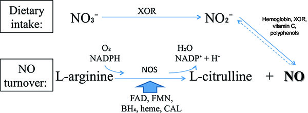
NO is produced from dietary sources via Nitrate-Nitrite-NO pathway or from endogenous turnover. The main differences between these pathways are basically the enzymes, the substrates, respectively nitrate and L-arginine, and the requirement of molecular oxygen (O2) for NO turnover pathway. Even though there are two processes, they are linked by the reduction of endogenously produced NO, which provides the largest endocrine source of directly bioavailable NO to inorganic nitrite (NO2) [15].

The NO synthesis from dietary intake is dependent on xanthine oxidoreductase (XOR). Among other functions, XOR is a major NO2 reductase enzyme linked to cellular NO signaling events [16-33] Fig. (1). This enzyme is essential for nitrate (NO3) use from diet.

Fig. (1). Physiological Nitric Oxide (NO) formation pathways. NO production using xanthine oxidoreductase (XOR) can be synchronized with nitric oxide synthase (NOS). NO turnover pathway is dependent on co-substracts as molecular oxygen (O2) and NADPH and cofactors such flavin adenine dinucleotide (FAD), flavin mononucleotide (FMN), tetrahydrobiopterin (BH4), heme and binding calmodulin (CAL) for NO synthesis.
OPEN IN VIEWER

In other way, NO produced from L-arginine requires an enzyme called nitric oxide synthase (NOS). There are three classical isoforms: endothelial (eNOS), neuronal (nNOS) and inducible (iNOS). These isoenzymes are derived from different genes and trigger diverse organic processes [34]. eNOS and nNOS are constitutively expressed and are dependent on Ca2+ for activation. In contrast, iNOS is usually expressed in proinflammatory processes and Ca2+ independent [35-43]. Besides the classical, there is a novel NOS isoform, mitochondrial NOS (mtNOS), which is present in mitochondria [44-46] and appears to regulate cellular oxygen consumption/energy metabolism without engendering oxidative stress [47, 48]. Positive vascular effects are well established as mediated by cellular pathways of NOS/L-arginine NO signaling [35, 49].

As almost all enzymes, NOS isoforms require cofactors. Tetrahydrobiopterin (BH4) is one of the critical cofactors for NOS activity. In conditions that will be approached in this article such as hypertension, BH4 is oxidized leading to NOS uncoupling [50, 51], increased Reactive Oxygen Species (ROS) and reduced NO production due to an electron flowing through the enzyme (Fig. 2).

3. NITRIC OXIDE INFLUENCES

NO acts on a number of protein targets through cell signaling. One of the most important physiological signaling is the activation of soluble guanylyl cyclase (GC) and the generation of cyclic guanosine monophosphate (cGMP) [9, 10, 52-55], especially for neurotransmission and vascular tonus functions. The transduction for the NO signaling is given by its reaction with superoxide anion (O2−•), resulting in NO inactivation and potent oxidant peroxynitrite (ONOO) formation. This compound causes oxidative damage, nitration, and S-nitrosylation of biomolecules including protein, lipids, and DNA [56, 57]. These damages are primordial to the development of the components predisposing MS and its symptoms, causing respectively hypertension and pain.
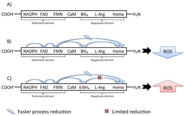
Fig. (2). NOS isoforms and reactive oxygen species (ROS) production through electron flowing. A) NOS enzyme schematic structure with main cofactors. B) Normal electron flowing in NOS enzyme: electron from flavin derived cofactors goes to Heme domain together with, but faster than, the electron transfer from BH4; however, before the next catalytic cycle can proceed, the BH4 has to be reduced. These oxidations lead to limitation of ROS production. C: electron flowing through NOS enzyme in hypertension: there is a disruption in BH4 oxidation, leading to NOS uncoupling, reduced no production and, consequently, increased ROS.
OPEN IN VIEWER

3.1. NO Pathway in Pain

The cardiovascular system functions reported on this article relies on blood circulation; namely, the association of angina and hypertension regarding nervous systems and blood circulation influenced by NO. In this sense, the expected NO functioning in the sensory perception of pain signaling and explanation are based on biochemical processes depicted on (Fig. 3).

The integrity of nervous system is essential for pain protective functions. Processes responsible for converting sensory stimuli to cellular transduction, enabling the recognition and characterization of the signal, modulate the frequency, rate and extent of the sensory perception of pain. Together with the NOS isoforms, glutamate receptor is involved in some of the important signaling processes [58]. Even though these proteins are reported to be associated to the regulation of sensory perception of pain, it is still unlinked, according to resources for “protein” search at the National Center for Biotechnology Information (NCBI) database [59], if they are positive or negative regulators or even part of the modulation of pain.

The usual NO pain pathway is showed on Fig. (3). However, how the NOS isoforms, mainly nNOS, are related to heart pain and the mechanism by which NO can sensitize the neuronal path to trigger the perception of pain remain partially unknown. Visceral and neuropathic are the main types of pain correlated to NO and ischemia, the principal cause of heart pain. Both pathways are nociceptors-sensitized on primary afferent C fibers, where the action potential is conducted to the central nervous and to secondary afferent neurons in spinal-cord dorsal horn. Then, the signal reaches areas of the brain responsible for localization and emotional aspects of pain, respectively, through spinothalamic and spinoreticular tracts. The main difference between visceral and neuropathic pain resides on the type of stimuli for which they respond to. Smooth muscle distension or contraction, capsule stretching surrounding an organ, ischemia, necrosis or inflammatory mediators trigger visceral pain; dissimilarly, the triggers for neuropathic pain pathway are trauma, surgery, diabetes mellitus, chemotherapy, radiotherapy, infection, malignancy and ischemia in which the damage occurs directly to central or peripheral nervous system [60].

Excluding nociception stimuli from extra cardiac issues, the main etiology for angina is ischemia. Intermittent ischemia in focal myocardial regions might result into functional alterations for both efferent and afferent cardiac adrenergic, and possibly vagal, nerve fibers. Additional mechanisms such as metabolic abnormalities might also adversely affect cardiac nerve fiber function [61, 62]. Cardiac stimuli are usually unable to elicit a painful response through afferent nerve fibers due to their low-sensitiveness; however, the fibers sensitivity to cardiac stimuli is increased if there are functional alterations, such the ones caused by ischemia. Therefore, the result is a painful response and consequent greater cardiac pain perception. This process is similar to cutaneous hyperalgesia due to peripheral sympathetic fiber injury described in literature [63]. Overall, impaired myocardial circulation generates ischemia stimulating the nociceptive pathway.

In order to elucidate the relationship between NO and angina, there are numerous studies with pharmacological approaches based on biotechnology researches applying knockout NOS mice [64-82]. Several studies have related drugs based on NO mechanisms and their influences on pain or ischemic signals.

Fig. (3). Nociceptive sensitization related to NO/ROS cascade. Neurotransmitter glutamate is secreted from the nociceptor terminal to the synaptic cleft and sensitizes AMPA receptors (AMPAr) on dorsal horn cells membrane. In long-term, the changes in membrane polarization affect the NMDA receptors (NMDAr). NMDAr sensitization allows the Ca+2 influx, which is essential to the neuronal Nitric Oxide Synthase (nNOS) activation. The nNOS activation is possible only if the inactive nNOS has all the cofactors (FAD, FMN, L-arginine and BH4) dimerized. For this, a lesion also increases the GTP cyclohydrolase (GCH1) levels, enhancing BH4. Ultimately, NO produced in the dorsal horn cells are released back into the synaptic cleft for closing guanyl synthase-induced K+ channels and for releasing Substance P. Respectively, the results are the opiate resistance in chronic pain and neural remodeling and hypersensitization.
OPEN IN VIEWER

In this sense, the development of NOS inhibitors was one of the first pharmacological approaches. Regarded as a therapy, since chronic pain patients showed a significant increase in NO plasma levels in comparison with healthy individuals [83], methylene blue (MB) is the most studied drug affecting NO mechanisms [64-73, 82]. MB directly inhibits constitutive and inducible NOS [65] through cGMP accumulation avoidance by GC enzyme blockage [65, 66]. A valuable property of MB is its antioxidant effects [66]; it acts inhibiting the formation of free oxygen radicals and O2−• by competing with molecular oxygen (O2). Therefore, the transfer of electrons by xanthine oxidase (XO) [68] is prevented. Studies have demonstrated MB decreasing pain levels in patients with chronic therapy-resistant neuropathic pain on the first 2 days after administration [69, 74].

Complementarily, studies using knockout mice analyzed NOS absence. In 2008, Nakata et al. [75] demonstrated NOS isoforms knockout mice in conditions of hypertension, hyperlipidemia, impaired glucose tolerance, insulin resistance, metabolic syndrome and presence of visceral obesity. In fact, targeted disruption of NOS genes leads to mutant mice development and allows a better understanding of NO mechanisms related to blood pressure regulation, endothelial dysfunction, response to vascular injury, response to stroke and cerebral ischemia, diet-induced atherosclerosis and cardiac contractility [76]. Results from such researches have shown the deletion of the eNOS gene led to increase blood pressure [84, 85]. Other studies analyzed the phenotype of nNOS knockout mice and noticed stomachs enlargement, several times bigger than normal size, demonstrating nNOS role in smooth muscle relaxation of the pyloric sphincter. nNOS knockout mice were also resistant to focal and global cerebral ischemia, consistent as a part of nNOS-derived NO function in cellular ischemic injury [76-80]. nNOS gene deletion has also been associated with more severe left ventricular remodeling after myocardial infarction [81].

3.2. NO Influences in Hypertension and Angina

According to data from the World Health Organization (WHO), cardiovascular diseases killed 17.5 million people in 2012, which are 3 in every 10 deaths globally distributed. Of these, 7.4 million people died due to ischemia and 1.1 million due to hypertensive heart diseases [86]. Both, ischemic and hypertensive heart diseases are directly influenced by coronary heart dysfunctions, which causes their usual symptom: angina [87]. In addition, one of the main prescriptions to patients with angina is glyceryl trinitrate, which belongs to the nitrates chemical group responsible for vasodilatation and consequent blood pressure decreasing.

There are many evidences on literature about the roles of NOS and cytochrome C (Cyt-C) in cardiovascular diseases [36, 75, 88-92]. The NOS influences on blood pressure and circulation vary depending on the type of the isoform. Nonetheless, Cyt-C is known to be part of NO production in strictly hypoxic conditions, such as ischemic angina. Despite these two well-studied proteins, there is a lack of evidence about what other factors are involved in such disrupted cardiovascular systems. In summary, it is possible only to correlate Cyt-C and NOS as part of the NO role in hypertension and angina.

The three NOS isoforms are active on cardiovascular system; however, the main enzymes related to hypertension and angina are eNOS and nNOS. iNOS contributes mostly to the pathophysiology of inflammatory diseases and septic shock [55, 75, 93].

NO produced by nNOS in nitrergic nerves is considered as a neurotransmitter responsible for stimulating NO-sensitive GC in its effector cells, thereby decreasing the tone of various types of smooth muscle including blood vessels [55, 94, 95]. nNOS functions include synaptic plasticity in the central nervous system (CNS), central regulation of blood pressure, smooth muscle relaxation and vasodilatation via peripheral nitrergic nerves [55]. Most importantly, nNOS plays a role in the regulation of vascular tone independent of effects from nNOS in the CNS [94, 95]. The blockage of nNOS activity in the medulla and hypothalamus causes systemic hypertension [96].

Complementarily, eNOS-derived NO dilates all types of blood vessels by stimulating soluble GC and increasing cGMP in smooth muscle cells [9, 10, 84]. NO from eNOS is a homeostatic regulator keeping blood vessels dilated, blood pressure, vasoprotection and anti-atherosclerotic effects. Although mostly expressed in endothelial cells, eNOS has also been detected in cardiomyocytes [94, 95]. Pharmacologically, vascular oxidative stress can be reduced and eNOS functionality restored with both renin- and angiotensin II- inhibitors and AT1 receptor blockers, and also with statins [55]. There are eNOS stimulators, the classic class of drugs for treating hypertension and myocardial infarction [35, 55, 97]. This choice for treatment is due to the powerful protective effect of eNOS-derived NO against the onset of atherogenesis. In short, NO from eNOS possesses the following effects: inhibition of platelet aggregation, vascular wall adhesion [98-100] and leucocyte adhesion to the vessel wall which are early events in the development of atherosclerosis; representing a critical factor for adaptive vascular remodeling to chronicle changes in blood flow [101]; controlling expression of genes involved in atherogenesis and angiogenesis post-ischemia [102]. The abrupt reduction on the bioavailability of eNOS-derived NO is observed after experimental myocardial infarction and in humans under heart failure condition [103, 104], contributing to impaired neovascularization [105]. Accordingly, endothelial NO can reduce the chances of angina episodes, once their effects are also correlated to ischemic-related angina.

Overall, nNOS and eNOS may have distinct roles in the physiological regulation of human microvascular tone in vivo [106]. Interestingly, low levels of nNOS have been shown in vascular smooth muscle cells as responsible to preserve some degree of vasodilatation when the predominant eNOS becomes dysfunctional [55, 107].

Hypertensive patients with metabolic syndrome and also patients with vascular diseases such as atherosclerosis show endothelial dysfunction due to reduced NO bioavailability and consequently impaired endothelium-dependent vasodilatation [108] associated with increased ROS production. There are several enzymatic systems potentially producing ROS in the vessels, including the Nicotinamide Adenine Dinucleotide Phosphate Hydrogen (NADPH) oxidases, XO mitochondrial respiratory chain, uncoupled eNOS [109] and nNOS [110-116]. Of these, NADPH oxidases are considered primary importance for ROS generation. Several isoforms of O2−•-producing NADPH oxidase are expressed in endothelial and smooth muscle cells, as well as in the adventitia layer [55].

The eNOS and nNOS produce large amounts of ROS when deprived of their critical cofactor BH4 or their substrate L-arginine [110-116]. An important stage of the electron transfer occurs in the Heme domain, which receives electron and enables oxygen binding. The reduction directly to the Heme domain is faster than Flavin reduction through the BH4 cofactor. Despite this faster process, the catalytic cycle can only proceed if BH4 was reduced. This difference leads to limitation of the reduced oxygen species productions by heme reduction. In hypertensive vessels Fig. (2), the disruption in electron flowing rather results in reduction of O2 at the prosthetic heme site than formation of NO [117]. The importance of the O2−• formation is due to the BH4 oxidation. Some in vivo studies [118] provided a mechanism for the predisposition to atherosclerosis suggesting NADPH oxidase as the initial source of ROS leading to BH4 oxidation. Endothelial and vascular smooth muscle cells-derived NADPH oxidase produces superoxide, respectively in early and advanced atherosclerosis stages [119]. Despite this knowledge, Laursen et al. [120] described ONOO as more potent BH4 oxidant than O2−• in hypertension. Indeed, myriad toxic effects of NO are recognized due to the subsequent generation of ONOO [121, 122] involved in inflammatory conditions [123, 124], neurodegenerative diseases [125, 126] and cardiovascular diseases [118, 120]. Therefore, NADPH oxidase- O2−• may not be an oxidant as relevant in hypertension as ONOO.

Similarly, the Cyt-C oxidase is a functionally competent ONOO reductase. It is suggested an enhanced NO production through a positive feedback mechanism for NO2-derived mitochondrial NO on a Cyt-C oxidase subunit. This protein recruitment is in state-dependent hypoxia; therefore, Cyt-C functional role is in hypoxic signaling events [127].

CONCLUSION

Manifold studies have proved or suggested the control and influences on blood pressure by NOS isoenzymes and have made correlation between NOS-NO and ischemic angina. This is because both, hypertension and ischemic angina, are part of a major MS that affect not just NO production by NOS, but also enzymes pathways, such Cyt-C, important to strictly anoxic conditions [16, 18, 128]. Due to this, it is still important to generate correlations between the many enzymes pathways already described on literature and angina for a better and more complete understanding of CVD.

In the vascular endothelium, BH4 mediates coupling of O2 reduction to heme-catalyzed L-arginine oxidation to form NO and L-citrulline [50]. In patients with MS, there is an inherently systemic inflammation and high risk of CHD [50]. The usual result is atherosclerosis in the coronary arteries leading to NADPH oxidase functioning and ROS products. Beyond ROS strengthen vascular lesions and NADPH oxidase functions, O2−• and ONOO oxide BH4. Even though the pathophysiologic control of endothelial BH4 levels in humans is poorly known, assembling the information described in the literature databases turns possible to have a better insight about the NO roles in cardiovascular symptoms such as angina.

LIST OF ABBREVIATIONS

BH4  = Tetrahydrobiopterin
cGMP  = Cyclic guanosine monophosphate
CHD  = Coronary heart disease
CNS  = Central nervous system
Cyt-C  = Cytochrome C
eNOS  = Endothelial - Nitric Oxide Synthase
GC  = Guanylyl cyclase
iNOS  = Inducible - Nitric Oxide Synthase
MB  = Methylene Blue
mRNA  = Messenger RNA
MS  = Metabolic Syndrome
mtNOS  = Mitochondrial - Nitric Oxide Synthase
NADPH  = Nicotinamide Adenine Dinucleotide Phosphate Hydrogen
NCBI  = National Center for Biotechnology Information
nNOS  = Neuronal - Nitric Oxide Synthase
NO  = Nitric Oxide
NO2  = Inorganic nitrite
NO3  = Nitrate
NOS  = Nitric Oxide Synthase
O2  = Molecular oxygen
O2−•  = Superoxide anion
ONOO  = Peroxynitrite
ROS  = Reactive Oxygen Species
XO  = Xanthine oxidase
XOR  = Xanthine oxidoreductase

AUTHORS’ CONTRIBUTIONS

Leticia Gonçalves and Carolina Baraldi Araujo Restini have made substantial contributions to conception and design and interpretation of material collected, as well as in drafting the manuscript, revising it critically for important intellectual content and have given final approval of the version to be published. Therefore, they have agreed to be accountable for all aspects of the work and ensuring questions related to the accuracy or integrity of any part of the work are appropriately investigated and resolved.

CONFLICT OF INTEREST

The authors declare that they have no significant competing financial, professional or personal influencing the performance or presentation of their work described in this manuscript.

ACKNOWLEDGEMENTS

The authors would like to thank to Dr. Reinaldo B. Bestetti and Dr. Suzelei de Castro França by the motivation to study the present aspects of NO pathways.

REFERENCES

1
Aboderin KA, Ben-Shlomo Y, Lynch JW, Yajnik CS, Kuh D, Yach D. Life Course Perspectives on Coronary Heart Disease, Stroke and Diabetes: Key Issues and Implications for Policy and Research. Geneva: Organization WH 2002. cited 2016 Mar 15
2
Kahn R, Buse J, Ferrannini E, Stern M. The metabolic syndrome: time for a critical appraisal: joint statement from the American Diabetes Association and the European Association for the Study of Diabetes. Diabetes Care 2005; 28(9): 2289-304.
3
Ford ES. Risks for all-cause mortality, cardiovascular disease, and diabetes associated with the metabolic syndrome: a summary of the evidence. Diabetes Care 2005; 28(7): 1769-78.
4
Galassi A, Reynolds K, He J. Metabolic syndrome and risk of cardiovascular disease: a meta-analysis. Am J Med 2006; 119(10): 812-9.
5
Gami AS, Witt BJ, Howard DE, et al. Metabolic syndrome and risk of incident cardiovascular events and death: a systematic review and meta-analysis of longitudinal studies. J Am Coll Cardiol 2007; 49(4): 403-14.
6
Ferrannini E. Metabolic syndrome: a solution in search of a problem. J Clin Endocrinol Metab 2007; 92(2): 396-8.
7
Schuman EM, Madison DV. A requirement for the intercellular messenger nitric oxide in long-term potentiation. Science 1991; 254(5037): 1503-6.
8
ODell TJ, Hawkins RD, Kandel ER, Arancio O. Tests of the roles of two diffusible substances in long-term potentiation: evidence for nitric oxide as a possible early retrograde messenger. Proc Natl Acad Sci USA 1991; 88(24): 11285-9.
9
Rapoport RM, Draznin MB, Murad F. Endothelium-dependent relaxation in rat aorta may be mediated through cyclic GMP-dependent protein phosphorylation. Nature 1983; 306(5939): 174-6.
10
Förstermann U, Mülsch A, Böhme E, Busse R. Stimulation of soluble guanylate cyclase by an acetylcholine-induced endothelium-derived factor from rabbit and canine arteries. Circ Res 1986; 58(4): 531-8.
11
Pantopoulos K, Hentze MW. Nitric oxide signaling to iron-regulatory protein: direct control of ferritin mRNA translation and transferrin receptor mRNA stability in transfected fibroblasts. Proc Natl Acad Sci USA 1995; 92(5): 1267-71.
12
Liu XB, Hill P, Haile DJ. Role of the ferroportin iron-responsive element in iron and nitric oxide dependent gene regulation. Blood Cells Mol Dis 2002; 29(3): 315-26.
13
Pozdnyakov N, Lloyd A, Reddy VN, Sitaramayya A. Nitric oxide-regulated endogenous ADP-ribosylation of rod outer segment proteins. Biochem Biophys Res Commun 1993; 192(2): 610-5.
14
Brüne B, Dimmeler S, Molina y Vedia L, Lapetina EG. Nitric oxide: a signal for ADP-ribosylation of proteins. Life Sci 1994; 54(2): 61-70.
15
Omar SA, Webb AJ. Nitrite reduction and cardiovascular protection. J Mol Cell Cardiol 2014; 73: 57-69.
16
Godber BL, Doel JJ, Sapkota GP, et al. Reduction of nitrite to nitric oxide catalyzed by xanthine oxidoreductase. J Biol Chem 2000; 275(11): 7757-63.
17
Li H, Samouilov A, Liu X, Zweier JL. Characterization of the magnitude and kinetics of xanthine oxidase-catalyzed nitrate reduction: evaluation of its role in nitrite and nitric oxide generation in anoxic tissues. Biochemistry 2003; 42(4): 1150-9.
18
Millar TM, Stevens CR, Benjamin N, Eisenthal R, Harrison R, Blake DR. Xanthine oxidoreductase catalyses the reduction of nitrates and nitrite to nitric oxide under hypoxic conditions. FEBS Lett 1998; 427(2): 225-8.
19
Godber BL, Doel JJ, Durgan J, Eisenthal R, Harrison R. A new route to peroxynitrite: a role for xanthine oxidoreductase. FEBS Lett 2000; 475(2): 93-6.
20
Maia LB, Moura JJ. Nitrite reduction by xanthine oxidase family enzymes: a new class of nitrite reductases. J Biol Inorg Chem 2011; 16(3): 443-60.
21
Golwala NH, Hodenette C, Murthy SN, Nossaman BD, Kadowitz PJ. Vascular responses to nitrite are mediated by xanthine oxidoreductase and mitochondrial aldehyde dehydrogenase in the rat. Can J Physiol Pharmacol 2009; 87(12): 1095-101.
22
Huang L, Borniquel S, Lundberg JO. Enhanced xanthine oxidoreductase expression and tissue nitrate reduction in germ free mice. Nitric Oxide 2010; 22(2): 191-5.
23
Jansson EA, Huang L, Malkey R, et al. A mammalian functional nitrate reductase that regulates nitrite and nitric oxide homeostasis. Nat Chem Biol 2008; 4(7): 411-7.
24
Zuckerbraun BS, Shiva S, Ifedigbo E, et al. Nitrite potently inhibits hypoxic and inflammatory pulmonary arterial hypertension and smooth muscle proliferation viaxanthine oxidoreductase-dependent nitric oxide generation. Circulation 2010; 121(1): 98-109.
25
Casey DB, Badejo AM Jr, Dhaliwal JS, et al. Pulmonary vasodilator responses to sodium nitrite are mediated by an allopurinol-sensitive mechanism in the rat. Am J Physiol Heart Circ Physiol 2009; 296(2): H524-33.
26
Baker JE, Su J, Fu X, et al. Nitrite confers protection against myocardial infarction: role of xanthine oxidoreductase, NADPH oxidase and K(ATP) channels. J Mol Cell Cardiol 2007; 43(4): 437-44.
27
McNulty PH, Scott S, Kehoe V, Kozak M, Sinoway LI, Li J. Nitrite consumption in ischemic rat heart catalyzed by distinct blood-borne and tissue factors. Am J Physiol Heart Circ Physiol 2008; 295(5): H2143-8.
28
Saraiva RM, Minhas KM, Zheng M, et al. Reduced neuronal nitric oxide synthase expression contributes to cardiac oxidative stress and nitroso-redox imbalance in ob/ob mice. Nitric Oxide 2007; 16(3): 331-8.
29
Tripatara P, Patel NS, Webb A, et al. Nitrite-derived nitric oxide protects the rat kidney against ischemia/reperfusion injury in vivo: role for xanthine oxidoreductase. J Am Soc Nephrol 2007; 18(2): 570-80.
30
Kelley EE, Batthyany CI, Hundley NJ, et al. Nitro-oleic acid, a novel and irreversible inhibitor of xanthine oxidoreductase. J Biol Chem 2008; 283(52): 36176-84.
31
Klocke R, Mani AR, Moore KP, Blake DR, Mapp PI. Inactivation of xanthine oxidoreductase is associated with increased joint swelling and nitrotyrosine formation in acute antigen-induced arthritis. Clin Exp Rheumatol 2005; 23(3): 345-50.
32
Webb A, Bond R, McLean P, Uppal R, Benjamin N, Ahluwalia A. Reduction of nitrite to nitric oxide during ischemia protects against myocardial ischemia-reperfusion damage. Proc Natl Acad Sci USA 2004; 101(37): 13683-8.
33
Berry CE, Hare JM. Xanthine oxidoreductase and cardiovascular disease: molecular mechanisms and pathophysiological implications. J Physiol 2004; 555(Pt 3): 589-606.
34
Aliciguzel Y, Ozen I, Aslan M, Karayalcin U. Activities of xanthine oxidoreductase and antioxidant enzymes in different tissues of diabetic rats. J Lab Clin Med 2003; 142(3): 172-7.
35
Stefano GB, Kream RM. Reciprocal regulation of cellular nitric oxide formation by nitric oxide synthase and nitrite reductases. Med Sci Monit 2011; 17(10): RA221-6.
36
Moncada S, Palmer RM, Higgs EA. Nitric oxide: physiology, pathophysiology, and pharmacology. Pharmacol Rev 1991; 43(2): 109-42.
37
Faraci FM, Heistad DD. Regulation of the cerebral circulation: role of endothelium and potassium channels. Physiol Rev 1998; 78(1): 53-97.
38
Kinoshita H, Tsutsui M, Milstien S, Katusic ZS. Tetrahydrobiopterin, nitric oxide and regulation of cerebral arterial tone. Prog Neurobiol 1997; 52(4): 295-302.
39
Cooke JP, Dzau VJ. Nitric oxide synthase: role in the genesis of vascular disease. Annu Rev Med 1997; 48: 489-509.
40
Stefano GB, Scharrer B, Smith EM, et al. Opioid and opiate immunoregulatory processes. Crit Rev Immunol 1996; 16(2): 109-44.
41
Fimiani C, Mattocks D, Cavani F, et al. Morphine and anandamide stimulate intracellular calcium transients in human arterial endothelial cells: coupling to nitric oxide release. Cell Signal 1999; 11(3): 189-93.
42
Stefano GB, Salzet M, Magazine HI, Bilfinger TV. Antagonism of LPS and IFN-gamma induction of iNOS in human saphenous vein endothelium by morphine and anandamide by nitric oxide inhibition of adenylate cyclase. J Cardiovasc Pharmacol 1998; 31(6): 813-20.
43
Moncada S, Palmer RM, Higgs EA. The discovery of nitric oxide as the endogenous nitrovasodilator. Hypertension 1988; 12(4): 365-72.
44
Kream RM, Stefano GB. Endogenous morphine and nitric oxide coupled regulation of mitochondrial processes. Med Sci Monit 2009; 15(12): RA263-8.
45
Brown GC. Nitric oxide and mitochondrial respiration. Biochim Biophys Acta 1999; 1411(2-3): 351-69.
46
Prevot V, Croix D, Rialas CM, et al. Estradiol coupling to endothelial nitric oxide stimulates gonadotropin-releasing hormone release from rat median eminence viaa membrane receptor. Endocrinology 1999; 140(2): 652-9.
47
Bates TE, Loesch A, Burnstock G, Clark JB. Mitochondrial nitric oxide synthase: a ubiquitous regulator of oxidative phosphorylation? Biochem Biophys Res Commun 1996; 218(1): 40-4.
48
Giulivi C, Kato K, Cooper CE. Nitric oxide regulation of mitochondrial oxygen consumption I: cellular physiology. Am J Physiol Cell Physiol 2006; 291(6): C1225-31.
49
Moncada S, Higgs A. The L-arginine-nitric oxide pathway. N Engl J Med 1993; 329(27): 2002-12.
50
Channon KM. Tetrahydrobiopterin: regulator of endothelial nitric oxide synthase in vascular disease. Trends Cardiovasc Med 2004; 14(8): 323-7.
51
Gangula PR, Sekhar KR, Mukhopadhyay S. Gender bias in gastroparesis: is nitric oxide the answer? Dig Dis Sci 2011; 56(9): 2520-7.
52
Furchgott RF, Cherry PD, Zawadzki JV, Jothianandan D. Endothelial cells as mediators of vasodilation of arteries. J Cardiovasc Pharmacol 1984; 6(Suppl. 2): S336-43.
53
Knowles RG, Palacios M, Palmer RM, Moncada S. Formation of nitric oxide from L-arginine in the central nervous system: a transduction mechanism for stimulation of the soluble guanylate cyclase. Proc Natl Acad Sci USA 1989; 86(13): 5159-62.
54
Garthwaite J. Glutamate, nitric oxide and cell-cell signalling in the nervous system. Trends Neurosci 1991; 14(2): 60-7.
56
Mikkelsen RB, Wardman P. Biological chemistry of reactive oxygen and nitrogen and radiation-induced signal transduction mechanisms. Oncogene 2003; 22(37): 5734-54.
57
Lee JH, Yang ES, Park JW. Inactivation of NADP+-dependent isocitrate dehydrogenase by peroxynitrite. Implications for cytotoxicity and alcohol-induced liver injury. J Biol Chem 2003; 278(51): 51360-71.
58
National Center for Biotechnology Information [Internet] U.S. National Library of Medicine, Bethesda MD: Protein; NOS isoforms; glutamate receptor. cited 2016 Mar 22. Available from: http://www.ncbi.nlm.nih.gov/ protein/144922606,1478510,148690957,148921954, 151555555, 197313638, 261278086, 261278088,2905806, 323635431,323635434,323635436,3355707,341942231, 41680705, 469067,475552,50403739, 545487902, 568936908, 568936910,568936912, 568936914, 5689
59
National Center for Biotechnology Information [Internet] U.S. National Library of Medicine Bethesda MD: Regulation of sensory perception of pain. Available from: http://www.ncbi.nlm.nih.gov/biosystems/512780 [cited 2016 Mar 22].
60
Reddi D, Curran N, Stephens R. An introduction to pain pathways and mechanisms [Internet]. London: University College London Hospital. Available from: https://www.ucl.ac.uk/ anaesthesia/StudentsandTrainees/ PainPathwaysIntroduction. [cited: 2016 Mar 23].
61
Lanza GA. Abnormal cardiac nerve function in syndrome X. Herz 1999; 24(2): 97-106.
62
Lanza GA, Crea F. The complex link between brain and heart in cardiac syndrome X. Heart 2002; 88(4): 328-30.
63
Baron R, Maier C. Reflex sympathetic dystrophy: skin blood flow, sympathetic vasoconstrictor reflexes and pain before and after surgical sympathectomy. Pain 1996; 67(2-3): 317-26.
64
Ashina M, Lassen LH, Bendtsen L, Jensen R, Olesen J. Effect of inhibition of nitric oxide synthase on chronic tension-type headache: a randomised crossover trial. Lancet 1999; 353(9149): 287-9.
65
Mayer B, Brunner F, Schmidt K. Inhibition of nitric oxide synthesis by methylene blue. Biochem Pharmacol 1993; 45(2): 367-74.
66
Mayer B, Brunner F, Schmidt K. Novel actions of methylene blue. Eur Heart J 1993; 14(Suppl I): 22-6.
67
Ohlow MJ, Moosmann B. Phenothiazine: the seven lives of pharmacologys first lead structure. Drug Discov Today 2011; 16(3-4): 119-31.
68
Salaris SC, Babbs CF, Voorhees WD III. Methylene blue as an inhibitor of superoxide generation by xanthine oxidase. A potential new drug for the attenuation of ischemia/reperfusion injury. Biochem Pharmacol 1991; 42(3): 499-506.
69
Peng B, Pang X, Wu Y, Zhao C, Song X. A randomized placebo-controlled trial of intradiscal methylene blue injection for the treatment of chronic discogenic low back pain. Pain 2010; 149(1): 124-9.
70
Licker M, Diaper J, Robert J, Ellenberger C. Effects of methylene blue on propofol requirement during anaesthesia induction and surgery. Anaesthesia 2008; 63(4): 352-7.
71
Salman AE, Salman MA, Saricaoglu F, Akinci SB, Aypar Ü. Pain on injection of propofol: a comparison of methylene blue and lidocaine. J Clin Anesth 2011; 23(4): 270-4.
72
Tan KY, Seow-Choen F. Methylene blue injection reduces pain after lateral anal sphincterotomy. Tech Coloproctol 2007; 11(1): 68-9.
73
Dudhgaonkar SP, Tandan SK, Kumar D, Naik AK, Raviprakash V. Ameliorative effect of combined administration of inducible nitric oxide synthase inhibitor with cyclooxygenase-2 inhibitors in neuropathic pain in rats. Eur J Pain 2007; 11(5): 528-34.
74
Miclescu AA, Svahn M, Gordh TE. Evaluation of the protein biomarkers and the analgesic response to systemic methylene blue in patients with refractory neuropathic pain: a double-blind, controlled study. J Pain Res 2015; 8: 387-97.
75
Nakata S, Tsutsui M, Shimokawa H, et al. Spontaneous myocardial infarction in mice lacking all nitric oxide synthase isoforms. Circulation 2008; 117(17): 2211-23.
76
Liu VW, Huang PL. Cardiovascular roles of nitric oxide: a review of insights from nitric oxide synthase gene disrupted mice. Cardiovasc Res 2008; 77(1): 19-29.
77
Zaharchuk G, Hara H, Huang PL, et al. Neuronal nitric oxide synthase mutant mice show smaller infarcts and attenuated apparent diffusion coefficient changes in the peri-infarct zone during focal cerebral ischemia. Magn Reson Med 1997; 37(2): 170-5.
78
Hara H, Huang PL, Panahian N, Fishman MC, Moskowitz MA. Reduced brain edema and infarction volume in mice lacking the neuronal isoform of nitric oxide synthase after transient MCA occlusion. J Cereb Blood Flow Metab 1996; 16(4): 605-11.
79
Huang Z, Huang PL, Panahian N, Dalkara T, Fishman MC, Moskowitz MA. Effects of cerebral ischemia in mice deficient in neuronal nitric oxide synthase. Science 1994; 265(5180): 1883-5.
80
Panahian N, Yoshida T, Huang PL, et al. Attenuated hippocampal damage after global cerebral ischemia in mice mutant in neuronal nitric oxide synthase. Neuroscience 1996; 72(2): 343-54.
81
Dawson D, Lygate CA, Zhang MH, Hulbert K, Neubauer S, Casadei B. nNOS gene deletion exacerbates pathological left ventricular remodeling and functional deterioration after myocardial infarction. Circulation 2005; 112(24): 3729-37.
82
Sim HL, Tan KY. Randomized single-blind clinical trial of intradermal methylene blue on pain reduction after open diathermy haemorrhoidectomy. Colorectal Dis 2014; 16(8): O283-7.
83
Farrokhi MR, Lotfi M, Masoudi MS, Gholami M. Effects of methylene blue on postoperative low-back pain and functional outcomes after lumbar open discectomy: a triple-blind, randomized placebo-controlled trial. J Neurosurg Spine 2016; 24(1): 7-15.
84
Ignarro LJ, Harbison RG, Wood KS, Kadowitz PJ. Activation of purified soluble guanylate cyclase by endothelium-derived relaxing factor from intrapulmonary artery and vein: stimulation by acetylcholine, bradykinin and arachidonic acid. J Pharmacol Exp Ther 1986; 237(3): 893-900.
85
Shesely EG, Maeda N, Kim HS, et al. Elevated blood pressures in mice lacking endothelial nitric oxide synthase. Proc Natl Acad Sci USA 1996; 93(23): 13176-81.
86
World Health Organization [Internet] The top 10 causes of death 2014 Available from: http://www.who.int/mediacentre/factsheets/fs310/en/ 2014. [cited 2016 Mar 23].
87
National Institutes of Health [Internet] Explore Angina: Department of Health and Human Services Available from: https://www.nhlbi.nih.gov/health/health-topics/topics/angina/causes 2014. [cited 2016 Mar 23].
88
Furchgott RF. The role of endothelium in the responses of vascular smooth muscle to drugs. Annu Rev Pharmacol Toxicol 1984; 24: 175-97.
89
Ignarro LJ. Biosynthesis and metabolism of endothelium-derived nitric oxide. Annu Rev Pharmacol Toxicol 1990; 30: 535-60.
90
Murad F. What are the molecular mechanisms for the antiproliferative effects of nitric oxide and cGMP in vascular smooth muscle? Circulation 1997; 95(5): 1101-3.
91
Shimokawa H. Primary endothelial dysfunction: atherosclerosis. J Mol Cell Cardiol 1999; 31(1): 23-37.
92
Bredt DS, Snyder SH. Nitric oxide: a physiologic messenger molecule. Annu Rev Biochem 1994; 63: 175-95.
93
Lange M, Enkhbaatar P, Nakano Y, Traber DL. Role of nitric oxide in shock: the large animal perspective. Front Biosci (Landmark Ed) 2009; 14: 1979-89.
94
Förstermann U, Closs EI, Pollock JS, et al. Nitric oxide synthase isozymes. Characterization, purification, molecular cloning, and functions. Hypertension 1994; 23(6 Pt 2): 1121-31.
95
Förstermann U. Regulation of Nitric Oxide Synthase Expression and Activity. In: Mayer B, Ed. Nitric Oxide. Berlin, Heidelberg: Springer Berlin Heidelberg 2000; pp. 71-91.
96
Toda N, Ayajiki K, Okamura T. Control of systemic and pulmonary blood pressure by nitric oxide formed through neuronal nitric oxide synthase. J Hypertens 2009; 27(10): 1929-40.
97
Landmesser U, Engberding N, Bahlmann FH, et al. Statin-induced improvement of endothelial progenitor cell mobilization, myocardial neovascularization, left ventricular function, and survival after experimental myocardial infarction requires endothelial nitric oxide synthase. Circulation 2004; 110(14): 1933-9.
98
Radomski MW, Palmer RM, Moncada S. The anti-aggregating properties of vascular endothelium: interactions between prostacyclin and nitric oxide. Br J Pharmacol 1987; 92(3): 639-46.
99
Busse R, Lückhoff A, Bassenge E. Endothelium-derived relaxant factor inhibits platelet activation. Naunyn Schmiedebergs Arch Pharmacol 1987; 336(5): 566-71.
100
Alheid U, Frölich JC, Förstermann U. Endothelium-derived relaxing factor from cultured human endothelial cells inhibits aggregation of human platelets. Thromb Res 1987; 47(5): 561-71.
101
Rudic RD, Shesely EG, Maeda N, Smithies O, Segal SS, Sessa WC. Direct evidence for the importance of endothelium-derived nitric oxide in vascular remodeling. J Clin Invest 1998; 101(4): 731-6.
102
Murohara T, Asahara T, Silver M, et al. Nitric oxide synthase modulates angiogenesis in response to tissue ischemia. J Clin Invest 1998; 101(11): 2567-78.
103
Bauersachs J, Bouloumié A, Fraccarollo D, Hu K, Busse R, Ertl G. Endothelial dysfunction in chronic myocardial infarction despite increased vascular endothelial nitric oxide synthase and soluble guanylate cyclase expression: role of enhanced vascular superoxide production. Circulation 1999; 100(3): 292-8.
104
Treasure CB, Vita JA, Cox DA, et al. Endothelium-dependent dilation of the coronary microvasculature is impaired in dilated cardiomyopathy. Circulation 1990; 81(3): 772-9.
105
Aicher A, Heeschen C, Mildner-Rihm C, et al. Essential role of endothelial nitric oxide synthase for mobilization of stem and progenitor cells. Nat Med 2003; 9(11): 1370-6.
106
Melikian N, Seddon MD, Casadei B, Chowienczyk PJ, Shah AM. Neuronal nitric oxide synthase and human vascular regulation. Trends Cardiovasc Med 2009; 19(8): 256-62.
107
Schwarz PM, Kleinert H, Förstermann U. Potential functional significance of brain-type and muscle-type nitric oxide synthase I expressed in adventitia and media of rat aorta. Arterioscler Thromb Vasc Biol 1999; 19(11): 2584-90.
108
Jeong SO, Son Y, Lee JH, et al. Resveratrol analog piceatannol restores the palmitic acid-induced impairment of insulin signaling and production of endothelial nitric oxide viaactivation of anti-inflammatory and antioxidative heme oxygenase-1 in human endothelial cells. Mol Med Rep 2015; 12(1): 937-44.
109
Mueller CF, Laude K, McNally JS, Harrison DG. ATVB in focus: redox mechanisms in blood vessels. Arterioscler Thromb Vasc Biol 2005; 25(2): 274-8.
110
Wever RM, van Dam T, van Rijn HJ, de Groot F, Rabelink TJ. Tetrahydrobiopterin regulates superoxide and nitric oxide generation by recombinant endothelial nitric oxide synthase. Biochem Biophys Res Commun 1997; 237(2): 340-4.
111
Xia Y, Tsai AL, Berka V, Zweier JL. Superoxide generation from endothelial nitric-oxide synthase. A Ca2+/calmodulin-dependent and tetrahydrobiopterin regulatory process. J Biol Chem 1998; 273(40): 25804-8.
112
Vásquez-Vivar J, Kalyanaraman B, Martásek P, et al. Superoxide generation by endothelial nitric oxide synthase: the influence of cofactors. Proc Natl Acad Sci USA 1998; 95(16): 9220-5.
113
Xia Y, Dawson VL, Dawson TM, Snyder SH, Zweier JL. Nitric oxide synthase generates superoxide and nitric oxide in arginine-depleted cells leading to peroxynitrite-mediated cellular injury. Proc Natl Acad Sci USA 1996; 93(13): 6770-4.
114
Pou S, Keaton L, Surichamorn W, Rosen GM. Mechanism of superoxide generation by neuronal nitric-oxide synthase. J Biol Chem 1999; 274(14): 9573-80.
115
Heinzel B, John M, Klatt P, Böhme E, Mayer B. Ca2+/calmodulin-dependent formation of hydrogen peroxide by brain nitric oxide synthase. Biochem J 1992; 281(Pt 3): 627-30.
116
Cosentino F, Lüscher TF. Tetrahydrobiopterin and endothelial nitric oxide synthase activity. Cardiovasc Res 1999; 43(2): 274-8.
117
Stuehr D, Pou S, Rosen GM. Oxygen reduction by nitric-oxide synthases. J Biol Chem 2001; 276(18): 14533-6.
118
Landmesser U, Dikalov S, Price SR, et al. Oxidation of tetrahydrobiopterin leads to uncoupling of endothelial cell nitric oxide synthase in hypertension. J Clin Invest 2003; 111(8): 1201-9.
119
Kawashima S, Yokoyama M. Dysfunction of endothelial nitric oxide synthase and atherosclerosis. Arterioscler Thromb Vasc Biol 2004; 24(6): 998-1005.
120
Laursen JB, Somers M, Kurz S, et al. Endothelial regulation of vasomotion in apoE-deficient mice: implications for interactions between peroxynitrite and tetrahydrobiopterin. Circulation 2001; 103(9): 1282-8.
121
Rodrigo J, Fernández AP, Serrano J, Peinado MA, Martínez A. The role of free radicals in cerebral hypoxia and ischemia. Free Radic Biol Med 2005; 39(1): 26-50.
122
Szabó C. Multiple pathways of peroxynitrite cytotoxicity. Toxicol Lett 2003; 140-141: 105-12.
123
Mabley JG, Liaudet L, Pacher P, et al. Part II: beneficial effects of the peroxynitrite decomposition catalyst FP15 in murine models of arthritis and colitis. Mol Med 2002; 8(10): 581-90.
124
Cuzzocrea S, Mazzon E, Di Paola R, et al. A role for nitric oxide-mediated peroxynitrite formation in a model of endotoxin-induced shock. J Pharmacol Exp Ther 2006; 319(1): 73-81.
125
Torreilles F, Salman-Tabcheh S, Guérin M, Torreilles J. Neurodegenerative disorders: the role of peroxynitrite. Brain Res Brain Res Rev 1999; 30(2): 153-63.
126
Kwak KH, Jung H, Park JM, et al. A peroxynitrite decomposition catalyst prevents mechanical allodynia and NMDA receptor activation in the hind-paw ischemia reperfusion injury rats. Exp Ther Med 2014; 7(2): 508-12.
127
Castello PR, Woo DK, Ball K, Wojcik J, Liu L, Poyton RO. Oxygen-regulated isoforms of cytochrome c oxidase have differential effects on its nitric oxide production and on hypoxic signaling. Proc Natl Acad Sci USA 2008; 105(24): 8203-8.
128
Li H, Samouilov A, Liu X, Zweier JL. Characterization of the magnitude and kinetics of xanthine oxidase-catalyzed nitrite reduction. Evaluation of its role in nitric oxide generation in anoxic tissues. J Biol Chem 2001; 276(27): 24482-9.
1800
1801
1802
1803
1804
Fig. (1).
Fig. (1). Physiological Nitric Oxide (NO) formation pathways. NO production using xanthine oxidoreductase (XOR) can be synchronized with nitric oxide synthase (NOS). NO turnover pathway is dependent on co-substracts as molecular oxygen (O2) and NADPH and cofactors such flavin adenine dinucleotide (FAD), flavin mononucleotide (FMN), tetrahydrobiopterin (BH4), heme and binding calmodulin (CAL) for NO synthesis.
Fig. (2).
Fig. (2). NOS isoforms and reactive oxygen species (ROS) production through electron flowing. A) NOS enzyme schematic structure with main cofactors. B) Normal electron flowing in NOS enzyme: electron from flavin derived cofactors goes to Heme domain together with, but faster than, the electron transfer from BH4; however, before the next catalytic cycle can proceed, the BH4 has to be reduced. These oxidations lead to limitation of ROS production. C: electron flowing through NOS enzyme in hypertension: there is a disruption in BH4 oxidation, leading to NOS uncoupling, reduced no production and, consequently, increased ROS.
Fig. (3).
Fig. (3). Nociceptive sensitization related to NO/ROS cascade. Neurotransmitter glutamate is secreted from the nociceptor terminal to the synaptic cleft and sensitizes AMPA receptors (AMPAr) on dorsal horn cells membrane. In long-term, the changes in membrane polarization affect the NMDA receptors (NMDAr). NMDAr sensitization allows the Ca+2 influx, which is essential to the neuronal Nitric Oxide Synthase (nNOS) activation. The nNOS activation is possible only if the inactive nNOS has all the cofactors (FAD, FMN, L-arginine and BH4) dimerized. For this, a lesion also increases the GTP cyclohydrolase (GCH1) levels, enhancing BH4. Ultimately, NO produced in the dorsal horn cells are released back into the synaptic cleft for closing guanyl synthase-induced K+ channels and for releasing Substance P. Respectively, the results are the opiate resistance in chronic pain and neural remodeling and hypersensitization.