All published articles of this journal are available on ScienceDirect.
Ultrasound-Guided Placement of a Renal Artery Stent Using an Intracardiac Probe for Transvascular Imaging
Abstract
In this set of images obtained during an experimental study using a porcine animal model, we introduce ultrasound guidance of percutaneous transluminal renal angioplasty and renal stenting. A state-of-the-art intracardiac ultrasound catheter is used here for transvascular scanning from within the lumen of the abdominal aorta, thus providing a field of view for navigation of a balloon catheter and a wire coil (“stent”) into each renal artery of a pig. This study is intended as a contribution to the growing field of minimally invasive interventions and their navigation by non-ionizing ultrasound imaging.
CASE REPORT
Percutaneous transluminal renal angioplasty and stenting have revolutionized treatment of renal artery stenoses [1]. These procedures are routinely done under fluoroscopic guidance, which can involve substantial radiation exposure. In this proof-of-concept experimental animal study, we demonstrate the feasibility of guiding stent placement by a 10F AcuNav ultrasound catheter (Siemens Medical, Mountain View, CA), which was connected through a SwiftLink cable to a Siemens Sequoia C256 ultrasound system. The AcuNav ultrasound catheter was primarily developed for intracardiac imaging applications, but has also been shown well suitable for transvascular imaging [2]. The purpose of this report was to introduce and illustrate navigation of renal artery stent placement by transvascular ultrasound imaging.
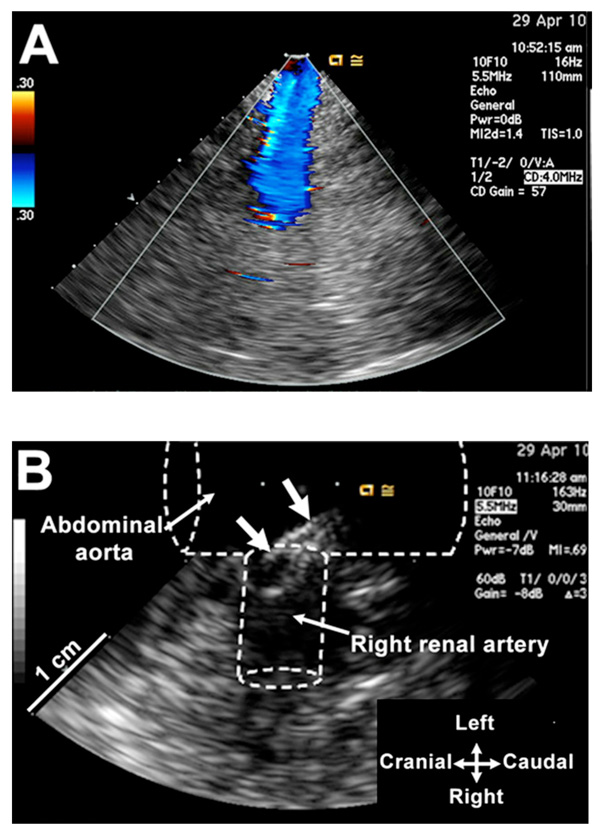
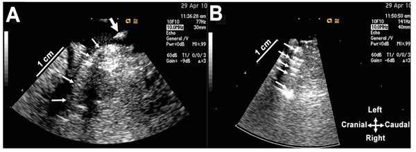
This study was approved by the Mayo Clinic Institutional Animal Care and Use Committee. An adult pig was anesthetized and the AcuNav ultrasound catheter inserted into the left femoral artery and advanced into the abdominal aorta until the kidney and renal artery Fig. (1A) on one side were identified. Then, an angulated-tip 8F Convoy catheter (EP Technologies, Boston Scientific, San Jose, CA) was inserted via the right femoral artery and advanced until its tip appeared within the ultrasound image. The tip was then easily guided into the renal artery orifice Fig. (1B). Subsequently, we inserted into the renal artery Fig. (2A) through the Convoy catheter lumen a PTCA catheter (Charger™; Cordis Corporation, Miami, FL) bearing a 23-gauge copper wire balloon-expandable coil-stent, as previously used experimentally [3]. We inflated and deflated the balloon, and removed

Color Doppler flow ultrasound image of the right renal artery obtained by the ultrasound catheter placed within the abdominal aorta. (B) Tip of the introducer catheter is entering into the right renal artery orifice (thick arrows). The drawing approximates the aortic and renal artery anatomy.

A balloon catheter with a coil on its tip has been placed into the right renal artery (small arrows) via the introducer catheter whose tip was subsequently pulled back (thick arrow). (B) The balloon catheter was inflated to deploy the coil, and after deflation pulled back from the renal artery lumen. The ultrasound scan shows reflections (arrows) from the coil.

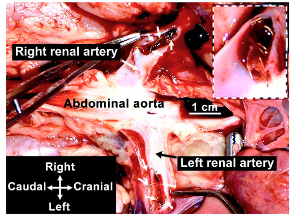
Postmortem photograph showing copper coils placed within right and left renal artery lumens. The insert is a close-up view of the coil inside the right renal artery just after dissecting its orifice.
both catheters, leaving the expanded coil deployed in the renal artery wall Fig. (2B). We repeated the procedure in the contralateral renal artery. At the end of the surgery, postmortem dissections documented successful coil placement inside each renal artery Fig. (3).
To our knowledge, this is the first transvascular ultrasound-guided placement of a renal artery stent by using an intracardiac ultrasound probe.
CONFLICT OF INTEREST
None declared.
ACKNOWLEDGEMENTS
We thank Naomi Gades, DVM, and Ronald Marler, DVM, PhD, for helpful discussions, Danielle Wright for secretarial work, and Ronald Buono, RDCS, for technical assistance. We are also grateful to Siemens Medical Solutions for loan of their SwiftLink cable for connection of our AcuNav catheter to a Siemens Sequoia C256 ultrasound system. Dr. Belohlavek was supported by NIH Grant EB009111.


